Get Healthy!
- Ernie Mundell and Robert Preidt
- Posted December 8, 2021
MRI Might Spot Concussion-Linked CTE in Living Patients
Right now, the devastating concussion-linked brain condition known as chronic traumatic encephalopathy (CTE) can only be diagnosed after death via autopsy. But new research could help change that, allowing doctors to someday spot the illness earlier.
According to the new study, MRI may be able to detect CTE while people are still alive.
"While this finding is not yet ready for the clinic, it shows we are making rapid progress, and we encourage patients and families to continue to participate in research so we can find answers even faster," said study senior author Dr. Jesse Mez. He directs Boston University's Alzheimer's Disease Center Clinical Core.
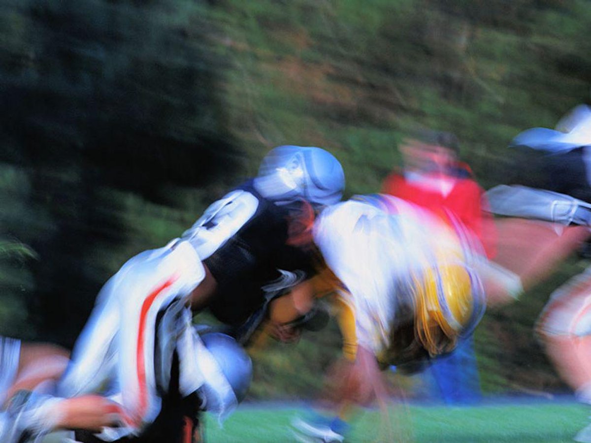
CTE has been linked with repetitive head impacts, and has been found in the brain autopsies of recently active and retired football players as well as other contact sport athletes. It's also been found in members of the military and victims of physical abuse.
Star NFL linebacker Junior Seau, who died by suicide in 2012, was later found to have suffered from CTE, as did the late football legend Frank Gifford.
Currently, it's only possible to diagnose CTE after death, but MRI may provide a way to identify the progressive brain disease sooner, the Boston research team said.
The investigators analyzed the medical records and MRIs of 86 deceased male brain donors. They included 55 men who'd been diagnosed with CTE after death and 31 men with healthy brains. The MRIs the men had had during their lifetimes were conducted an average of four years before their deaths.
Compared to those with healthy brains, the MRIs showed that the men diagnosed with CTE after death had shrinkage in key regions of the brain associated with CTE, as well as other abnormalities.
"Specifically, those with CTE had shrinkage in the frontal and temporal lobes of the brain, the regions most impacted by CTE," Mez said in a university news release.
According to study lead author Michael Alosco, "MRI is commonly used to diagnose progressive brain diseases that are similar to CTE such as Alzheimer's disease. Findings from this study show us what we can expect to see on MRI in CTE. This is very exciting because it brings us that much closer to detecting CTE in living people." Alosco is associate professor of neurology at the Boston University School of Medicine and co-director of the Alzheimer's Disease Center Clinical Core.
Speaking in the news release, Alosco said that "there is more to do as we still need to understand whether the patterns we saw on MRI are specific to CTE, that is, do they differentiate CTE from Alzheimer's disease and other causes of dementia."
Dr. Andrew Rogove directs stroke care at Northwell Health's South Shore University Hospital in Bay Shore, N.Y. He wasn't involved in the new research, but called it "a very interesting beginning into the possibility of diagnosing CTE among the alive patient."
Rogove said that would be a boon to patients, and "may eventually lead to identification and development of biomarkers that can make earlier diagnosis possible."
The study was published online Dec. 7 in the journal Alzheimer's Research & Therapy.
More information
The Concussion Legacy Foundation has more on CTE.
SOURCES: Andrew D. Rogove, MD, PhD, DPN-N, stroke director, Northwell Health's South Shore University Hospital, Bay Shore, N.Y.; Boston University, news release, Dec. 7, 2021


